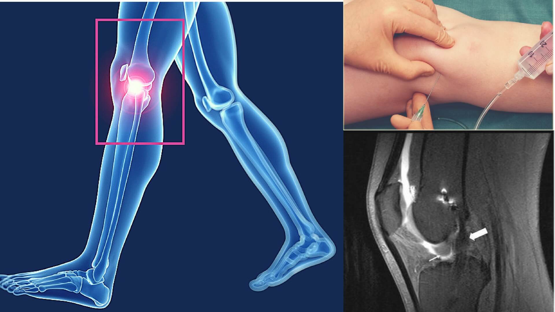
Arthrogram in layman’s language can be described as X-ray of joints that produces multiple pictures. The procedure is different than ordinary X-ray as arthrogram requires injection of a contrast material like dye. Anatomical structure of the joint that comprises of ligaments, tendons, cartilage, muscles and joint capsule are captured clearly on paper by arthrogram which normal X-ray is unable to do. The series of pictures thus obtained are used by orthopedics to diagnose joint disorders and decide treatment approach. Sometimes, arthrogram can be used in combination with MRI or CT if arthrogram pictures provide inadequate information. Arthrogram can be carried out on various joints, such as knee joint, shoulder joint, ankle joint, elbow joint, jaw and wrist joint.
Let’s understand arthrogram in detail.
Continue reading “This Post Will Make You Know About Arthrogram: Read Or Miss Out”