What is MRI Glioblastoma?
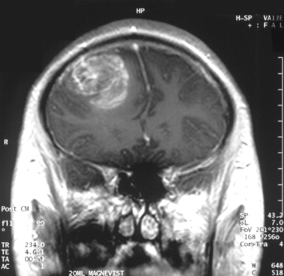
MRI Glioblastoma is the investigative technique used to study and diagnose brain cancer affecting the supporting cells of human brain.
Magnetic Resonance Imaging (MRI) is an advanced technique which studies internal organs of a patient using magnetic field. MRI studies involve generating a magnetic field of specific intensity and focusing it over that part of patient’s body which is to be studied. The magnetic waves interact with hydrogen particles present within patient’s body, which results in generation of radio waves. Interaction between radio waves and magnetic waves leads to formation of images which can be seen on a computer screen and used to diagnose various health conditions.
MRI of brain requires to be done in order to diagnose or study glioblastoma. A normal MRI suffices often, but in case this fails to provide adequate information about the glioblastoma, specialized MRI study is needed. Functional MRI may be required in such cases.
Functional MRI:
Functional MRI involves a scan while a person is performing a particular activity viz. walking and alternatively while the person is resting. This is done to note which area of brain is stimulated during the patient’s activity.
How is MRI Glioblastoma performed?
Patient has to visit a hospital or diagnostic center for getting MRI Glioblastoma done. Since this investigation requires great skill and precision, it is advisable to visit a reputed diagnostic center. Following are the steps involved in performing the procedure:
Patients are asked to change their clothes and dress up in a clean sterile gown provided by the diagnostic center.
All jewelery and accessories have to be removed while the scan is being performed.
Patients who have pacemaker, orthopedic screws, prosthetic joint, chemoport device, arterial stents fitted in their body, should notify the doctor before MRI begins.
Expecting mothers have to inform doctor about their pregnancy prior to the scan.
Before the scan, a heavy dye may be injected into the patient’s blood through a vein in the arm. This dye gets absorbed into the brain tissue, making the cancerous portion stand out more clearly.
Patient is made to lie on a flat bed which is slided inside a hollow wide tube (closed MRI) or between two large magnetic plates (open MRI).
A magnetic field of predetermined intensity is generated and focused on the patient’s skull. The scanner will study and display images of the patient’s brain from various axes and angles.
The entire process takes about 45 minutes to one and a half hour. After the procedure is completed, the patient may go home immediately.
Sometimes, glioblastoma causes the patient to experience epileptic seizures (fits). Such an attack during the scan may hamper the scan results. Such patients might require anesthesia to keep them still and calm during the procedure.
Indications of MRI Glioblastoma:
Primary Diagnosis: MRI brain may be ordered as a first line investigation to diagnose glioblastoma when no other investigations have been done previously.
Second line of investigation: It may also be required for those patients who have undergone previous investigations for glioblastoma, but the diagnosis has not been clear, or it has not provided adequate information about extent and intensity to which brain cancer has spread.
Follow up: For patients of glioblastoma who are undergoing treatment, MRI is a good tool to study how effective the treatment is and whether the patient is recovering in the expected manner.
Guided biopsy: MRI scan can be performed for glioblastoma when biopsy of the cancerous brain tissue is to be performed. MRI scan allows the pathologist and neurosurgeon who are performing the biopsy, to visualize the tumor location precisely.
MRI Glioblastoma is a time consuming study requiring immense precision and skill. Patients should follow all pre and post procedures instructions carefully to achieve the best scan results.