Radiology is the branch of medical studies that deals with radiation usage for diagnosing and treating diseases afflicting human beings. Radiology is an important part of treatments as it reveals complications and causes of disorders in the body. The doctor is then able to decide on the course of treatment to be given to his patient. 
Have you ever wondered what role the radiologists or laboratory technicians have in diagnosing the bug in your tummy as something more serious? Not many people would ponder on this and the uninteresting gargantuan setup of equipment in a laboratory.
Read on to find out what actually these radiological tests done by radiologists and doctors are all about…
What are radiological tests?
Radiological tests are a bunch of imaging and scanning tests meant for detecting abnormalities and anomalies affecting general well-being and good health in people. Through imaging, the doctor gets a view of the inner anatomy in human beings for better understanding of his/her disease.
What are the types of radiology?
Diagnostic & Therapeutic Radiology are the 2 sub-specialties of radiology.
- Diagnostic Radiology
Diagnostic Radiology employs imaging techniques for diagnosis of diseases and subsequent treatment ahead. Interventional Radiology is one sub specialty
belonging to diagnostic radiology discipline. Interventional Radiology makes use of minimally invasive techniques of diagnosing the patient using procedures which require less pain and faster recovery time.
In Interventional Radiology, radiological image guiding techniques like ultrasound, CT or MRI scan methods for treatment are used. Treatment of blood clots formed in lungs through device placement is done by interventional radiologists. Similarly, blood clots in veins and varicose veins are also treated using interventional radiology techniques which are relatively safe and require less of time in the hospital. - Therapeutic Radiology
Therapeutic Radiology involves radiation therapy methods for treating diseases like cancers. Radiation therapy generally is used for killing and minimizing growth of cancerous cells in the body. It is used to destroy malignant cells and also used immediately after adjuvant therapy for targeting remnant cancerous cells if any.
What are the types of radiological tests for patients?
There are many radiological tests useful for diagnostic purposes ordered by doctors. Some of them are as listed below:
- X-ray
- Magnetic Resonance Imaging (MRI)
- Magnetic Resonance Angiogram (MRA)
- Ultrasound
- Color Doppler
- Computed Tomography (CT Scan)
- Mammography
- PET Scan
- Bone Density test
- Biopsy
- Fluoroscopy
- X-ray
This is the most common and ubiquitous form of radiological testing. It is a type of imaging technology which helps the doctor to see internal structure of a patient.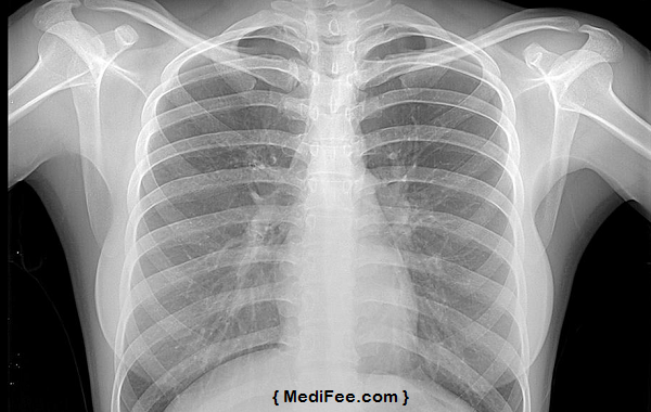
These days, X-ray imaging is even ordered by dentists before initiating line of treatment for dental worries. Apart from dentists, X-rays are helpful for cardiologists in examining blocked blood vessels. Orthopedic specialists make use of X-rays for finding causes of osteoporosis and managing fracture cases. X-ray is also used for diagnosis of bone cancer and helps in deciding bone cancer treatment procedure. An abdomen X-ray enables the doctor to examine stomach, kidneys, intestines and spleen when there is a sudden bout of stomach ache or vomiting in patients. - Magnetic Resonance Imaging (MRI)
As the name suggests, this imaging makes use of magnetic technology and radio waves for creating images of the body. MRI scans are used for detecting tumors in brain, brain injuries, dementia, multiple sclerosis etc. They are also done to ascertain spinal tumors, fractures, reproductive system issues, cysts in urinary tract too. You can find the prices of MRI scan in various Indian cities here. - Magnetic Resonance Angiogram (MRA)
Magnetic Resonance Angiogram provides anatomical and very minute information which isn’t possible even using ultrasound, CT scan or X-ray imaging. Calcium deposits and fat buildup in blood vessels leading to brain, narrowing of blood vessels which provide blood to legs, heart and kidneys, can be detected using Magnetic Resonance Angiogram. A bulging of any of the blood vessels supplying blood is seen to be harmful and calls for surgery. - Ultrasound
Ultrasound, also known as sonography, makes use of sound waves for producing pictures of concerned internal organs in the body. It is used for tracking growth of infants in pregnant women. Since ionizing radiation is not used, this is extremely safe. Ultrasound helps cardiologists in identifying aftermath and damage to one’s heart in event of a heart attack. A breast ultrasound enables oncologists to detect lumps and tumors in breasts. - Color Doppler
Color Doppler or Doppler Ultrasound detects blood flow through major arteries and veins in legs, arms and neck. Deep vein thrombosis which forms blood clots in veins present in legs is identified with this ultrasound scan. Unborn babies’ growth and development is tracked using this test too. Arterial plaque which can trigger a stroke can be detected using Color Doppler. - Computed Tomography (CT Scan)
Computed Tomography is a type of imaging technology for generating pictures of blood vessels, vein bones, soft tissues etc. The CT scanner generates 3D images of the entire body during scanning. The images are much more comprehensive than those generated from an X-ray scan. CT scans are used for detecting tumors, cancers, kidney stones etc. It is the best imaging modality for detecting abdominal ailments, sinusitis, spine diseases, chest pain etc. This type of imaging also lets women know if they are pregnant. - Mammography
Mammography involves screening of breasts for possible tumors that could possibly be breast cancer lumps. Several X-rays of either breasts are taken.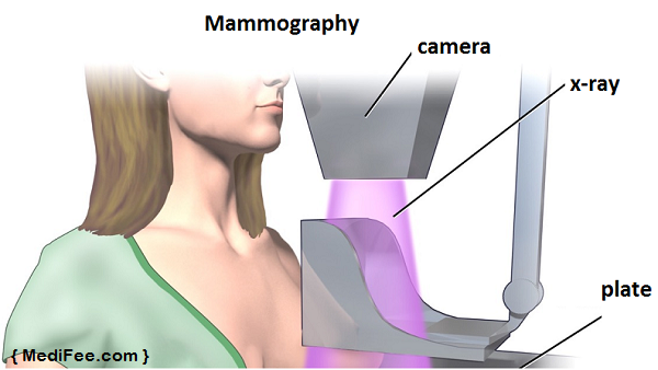 The X-ray images are transferred to a computer by the digital mammogram to help radiologist in further analysis. The images help gynecologists to detect lumps, cancerous and non cancerous growths and calcium deposits inside breasts.
 - PET CT Scan
PET Scan is ordered by doctors for detecting brain and heart diseases and different cancer types. Radioactive tracers are administered to the patient through an injection or a drink. The tracers take about an hour before showing effect. After which the patient is made to slide inside the PET machine. Drinking plenty of liquids for eliminating the tracers out of your system is essential. - Bone Density test
Bone Density tests make use of X-rays for ascertaining osteoporosis in a person.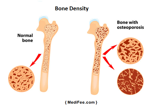 The test usually lasts 10-15 minutes and is done using a central machine for passing radiation. Find prices for bone density test near your city, here.
 - Biopsy
Biopsy is done either after extracting a small amount of tissues or used in combination with imaging modalities like X-ray, ultrasound, MRI etc. Cancerous and non cancerous growths can be diagnosed using a biopsy. Biopsies are of different variety such as, abdominal, endometrial, liver, muscle, testicular etc. - Fluoroscopy
Fluoroscopy is an imaging procedure used in conjunction with other diagnostic procedures. It is used for viewing the internal structure or a particular organ in the body on a monitor.
So, here was a short overview of what exactly radiological tests are. Doctors advising patients to undergo these tests for diagnosis is becoming more and more common. People turning to the web for getting basic, but useful information, is also soaring. It thus is better to know their nitty-gritties before undergoing tests for having better clarity of the same.