The Magnetic Resonance Imaging (MRI) is a unique technique which uses magnetism, radio waves and a computer to manipulate properties of water molecules (mostly H+ ions) in our bodies to produce an image.
The H+ ions (protons) possess paramagnetic properties because of which they easily align themselves in a peculiar fashion in presence of a magnetic field. Once the patient is inside the scanner, radio waves and magnetic resonance (MR) together create a variable environment which alters the ‘spin’ of protons after absorption of energy. The field is then switched off during which protons return to their normal energy state, known as precession. The radio signals produced in this state are picked by scanners which finally produce an image.
Result?? A detailed picture of both the hard and soft tissues with the benefit of no harmful/non-ionizing radiations.
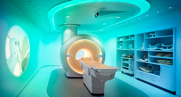
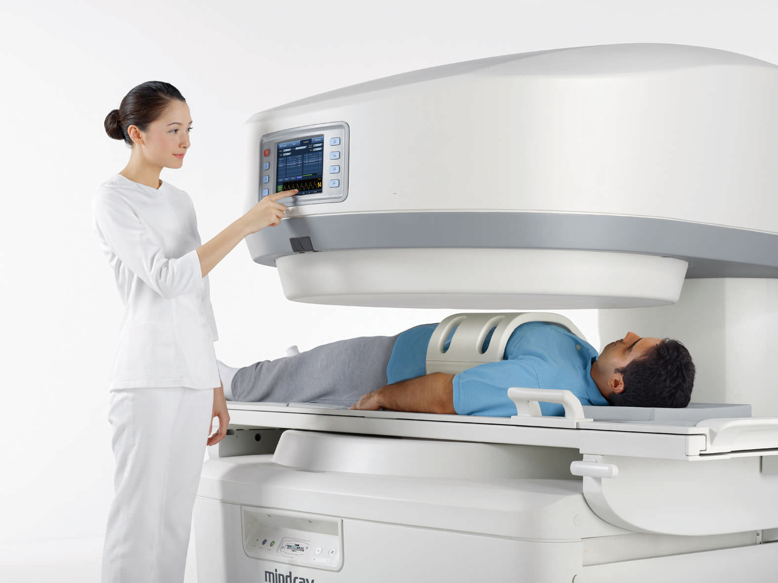
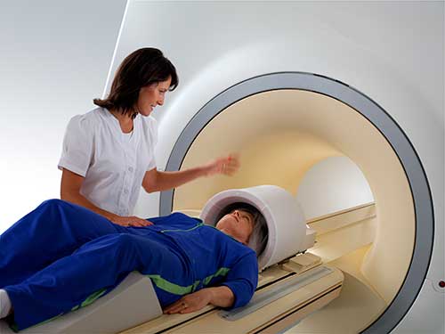
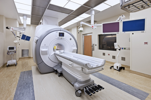
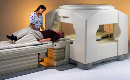
In routine settings, an MRI machine is a hollow cylindrical structure in which the patient has to stay for sometime (in a lying down position) till the image is taken. A magnetic strength of about 0.2-3 Tesla is applied for 90 minutes, or more.
Types of MRI:
MRI scans can be classified in a variety of ways depending on its construction, properties and applications. Here we will go through a few of them to understand their similarities and differences.
Based on Construction:
MRI designs have been improved upon to provide patients a greater degree of comfort and decreasing the chances of claustrophobia.
-
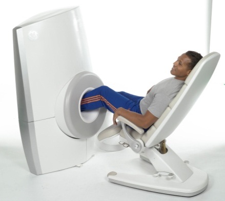
Open MRI –
Open-MRI-Machine
For patients who may feel uncomfortable in enclosed spaces (claustrophobhia), this is a very convenient option. An open MRI can have a wider bore diameter (more than 60 cm) or be open on the sides. The patient will still have to inside on a sliding table, but the process becomes more comfortable.
The only downside here is unlike a closed MRI image details are less clear because of inability to contain the magnetic field as desired.
-
Extremity MRI –
Extrimity-MRI-Machine
Similar to a closed MRI, it will study the limbs in detail. The difference here is that it is specially customized for our extremities (arms & legs), meaning a smaller scanner, minimal restriction on movements and no claustrophobia.
This is useful in diagnosing stress injuries, arthritis, infections, tumors, soft and hard tissue injuries, etc.
-
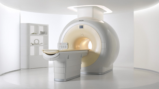
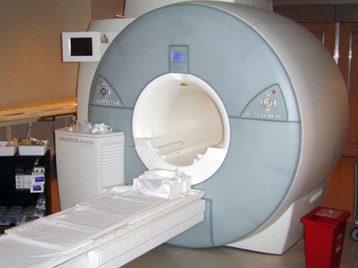
3 Tesla MRI –
3-Tesla-MRI-Machine
As compared to a standard MRI, this has the ability to produce a magnetic field double the conventional value. Thus, detailed images can be created in a lesser period of time.
Blood vessels as small as 200-300 micron can be easily visualized and vascular problems anywhere in the body can be identified. A magnetic field of this strength can catch developmental anomalies in the brain which are poorly visualized in a 1.5 Tesla MRI.
Better sound control while imaging is seen in this MRI.
Based on Properties & Applications:
Keeping in mind the requirements and physiology of different body systems, MRI scans are now ‘customized’ as needed. Let us take a look at the various types:
-
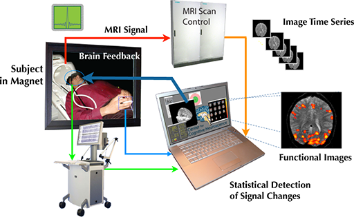
Functional MRI (FMRI):
Functional-MRI-Machine
Unlike the conventional MRI scans that create images of organs, an FMRI looks at how blood flows within the nervous system.
An adult uses about 20% of the body’s oxygen, where the active neurons (brain cells) use more of it. The image is made using variations in neural activity because of BOLD – blood oxygen level dependent effect, which is dependent on the amount of deoxyhemoglobin present. When neurons become active, cerebral blood flow increases in that particular region of the brain. This increases the amount of oxyhemoglobin with respect to that of deoxyhemoglobin in the area.
Oxyhemoglobin possessing diamagnetic properties doesn’t interfere much with the magnetic resonance signal. Deoxyhemoglobin, on the hand is paramagnetic and dephases (puts out of synchronization) the magnetic signal generated by MRI machine. More the deoxyhemoglobn, lesser the amount of interfering signal.
This logic is applied very efficiently in diagnosing the effects of stroke and degenerative diseases like Alzhiemer’s on the brain. Along with this, studying the effects of blood flow in the brain using during brain surgery helps in identifying vital areas affecting thought process, speech etc. hence guiding the surgeon where to exercise utmost care and caution.
-
Interventional MRI:
Interventional-MRI-Machine
With its low dose radiation, MRI can be of great help while performing minimally invasive surgeries, to assess the safety and correctness of the procedure being performed. Interventional MRI finds great use in biopsies of suspected lesions, to remove tissues non-invasively (thermal ablation), various heart procedures, etc. Interventional MRI importance is great in neurosurgical procedures offering greater depth and detailing of images.
However care should be taken that these procedures are performed without the use of ferromagnetic instruments (not have any ‘magnetic’ properties) such as titanium and other MR (magnetic resonance) friendly instruments.
-
Cardiac MRI:
Cardiac-MRI-Machine
Technically known as cardiovascular magnetic resonance (CMR), it operates on the same principles as that of an MRI, but customized for use in ‘matters of the heart.’ Anatomy of the heart, cardiomyopathies, pericardial diseases, coronary artery disease, thrombus and ventricular disorders can be easily diagnosed using this technique. The CMR will be carried out in a fashion similar to the conventional MRI, but you may have to hold your breath for short periods of time when image will be recorded.
During this procedure, ECG leads are attached to your body to provide electrical records of the heart in synchronization with the images produced, hence providing a quick and comprehensive information to your doctor.
It has many advantages such as a clear image quality, accuracy, low-energy radiation and safety. Most importantly, it can be easily correlated with an ECG at the same time.
Sadly, the availability of this technology is an issue owing to high MRI scan costs and dearth of skilled technicians well versed with its use.
-
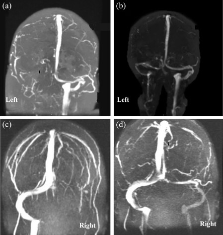
Magnetic Resonance Angiography (MRA):
Magnetic-Resonance-Angiography-Machine
Angiography in general is a medical test which s used to diagnose abnormalities in blood vessels of the body, most commonly the arteries of the brain, legs, neck, chest, abdomen, pelvis and arms . An angiogram can be obtained using fluoroscopy, computed tomography or an MRI scan.
There are two types of MRA:
-
-
-
-
Flow dependent –
This relies on the flow of blood in the body. Diagnosis of any abnormality is based on the comparison of blood flow in static tissue and vessels. A contrast medium is generally used.
-
Flow independent –
Beneficial for patients having slow blood flow. Here, the diagnosis is made independent of the flow of blood, rather it is based on T1, T2 and chemical shifts of different tissues.
-
-
-
2D and 3D images are made possible with the help of orienting the MR at different angles.
The contrast media, if used generally contains gadolinium which helps in producing brighter images of the vasculature. And for those who are allergic to contrast media, flow independent MRA is the best solution nullifying any risk of hypersensitivity reactions.
-
Magnetic Resonance Venography (MRV):
Brain-magnetic-resonance-venography
An experimental technique of looking into veins but promising enough to offer accurate results, MRV checks the flow of blood in veins. Strokes, aneurysms, vascular disease or any obstruction in veins can be detected using this method. A contrast media (a dye that makes your blood vessels look opaque in the scan) may or may not be injected into the veins.
Evaluating veins using this method is a less commonly used approach due to the availability of better investigative techniques such as the Doppler angiography.
-
Diffusion MRI:
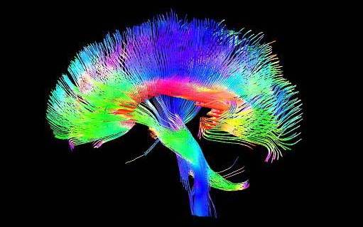
DTI-of-white-matter-in-the-brain
Diffusion is defined as the movement of molecules from a region of high concentration to a lower one. Water molecules in the body follow the same laws of diffusion too. In the human body, water molecules behave in an anisotropic fashion, i.e. their motion is direction dependent. It will follow a specific pattern.
This principle is used to determine the direction of nerve fibres in the brain, especially white matter. Combined with diffused tensor imaging (DTI), diffusion can be studied in multiple directions to create a brain map showing connectivity in different regions of the brain. Stroke can be easily detected using this method.
-
Magnetic Resonance Spectroscopy:
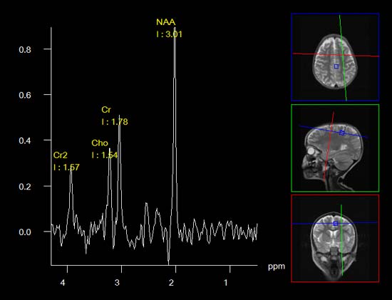
Also known as nuclear magnetic resonance spectroscopy (NMR), this technique operates by detecting changes in metabolites produced in response to certain diseases, especially that of the nervous system and muscular system. Where an MRI scan creates a 2D image of the concerned area, an MRS will supplement information using 1H signals to compare the chemical compositions of normal tissue versus abnormal tissues (tumors, stroke, epilepsy, etc.)
An MRS calculates concentration of H+ ions produced in the tissues along with other metabolites as alanine, choline, creatine, choline, N-acetyl aspartate, lipid, lactate, etc. in concentrations of parts per million (ppm). These concentrations are then plotted on a graph which are then compared by a clinician for appropriate diagnosis.
-
Real Time MRI:
This technique creates a ‘live’ imaging of the structures to be examined. This has special benefits when it comes to recording blood flow in the heart, or recording joint kinetics of temporomandibular joint, knees and wrists. Movements of lips, tongue and soft palate while speaking (articulatory phonetics) or swallowing can be recorded using this method.
-
Magnetic Resonance Guided Focused Ultrasound:
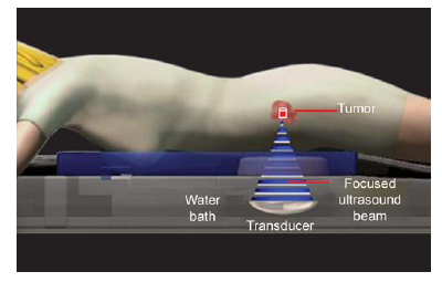
Magnetic-Resonace-Guided-Focused-Ultrasound
A place where ultrasound meets image precision of the MRI.
Here non-invasive ultrasound beams are used in the frequency of 220 KHz to 680 KHz to generate heat upto 85 degrees centigrade. An MRI scanner has the unique property of providing accurate image details along with detecting temperature changes. This enables the surgeon to use controlled heat generated by ultrasound waves to perform the required surgery/diagnostic procedure.
-
Multinuclear Imaging:
Multinuclear-Imaging
Presently, MRI scans can detect differences in ‘spin’ of H+ atoms (protons) or activity of water molecules in the body. A multinuclear MRI will widen its scope of detection to other atoms which can produces a net nuclear spin. Research is being done on nuclei such as helium-3, lithium-7, sodium-23, potassium-39, oxygen-17, etc. because of their important role in many biological processes.
Come to think of it, an oxygen-17 MRI can be used to detect oxygen consumption in a non-invasive way. Different spins of different atoms can be used to generate high quality images with greater degree of contrasts. Hyperpolarized He3+ if inhaled and then scanned for inside the chest cavity can help in locating condition of air spaces in the lungs.
With dedicated research and execution, multinuclear imaging has the potential to indicate varying bone densities or studying the distribution of certain therapeutic elements in the brain to aid in potential drug therapy.
-
Molecular Imaging Using MRI:
Our bodies produce certain biomarkers (gene, molecule) when it suffers from any pathology. These biomarkers if detected early can help in prompt treatment and stop further progression of the disease. But detecting these markers with an imaging modality requires high degree of sensitivity & specificity at room temperatures. To deal with this challenge, research is being conducted on MRIs to identify targeted specific contrast agents during recording of image which at the same time may serve as a vehicle for drug delivery to the affected site.
Apart from these types, there are some specialized features that have been incorporated to optimize the output of MRI scans:
-
Magnetization Transfer MRI
-
T1rho MRI
-
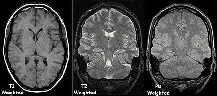
Proton Density Weighted
-
Fluid Attenuated Inversion Recovery (FLAIR)
-
Susceptibility Weighted Imaging (SWI)
-
Neuromelanin Imaging
MRI scans have revolutionized the way images are perceived. With its dual benefit of high quality images with no radiation exposure, abnormalities and disorders can be identified easily, thus ensuring prompt delivery of appropriate treatment. Every small step in research is being dedicated to make MRI scans a fast imaging technique with a wider scope of activity.
What type of MRI is used for nerves brain tumour and blood cloth