Everyone would like to be in the ‘Top 5’ or ‘Top 10’ of things. Sadly, when it comes to diseases, nobody would like to rank them, even in the ‘Most deadly’ or ‘Highly morbid’ categories.
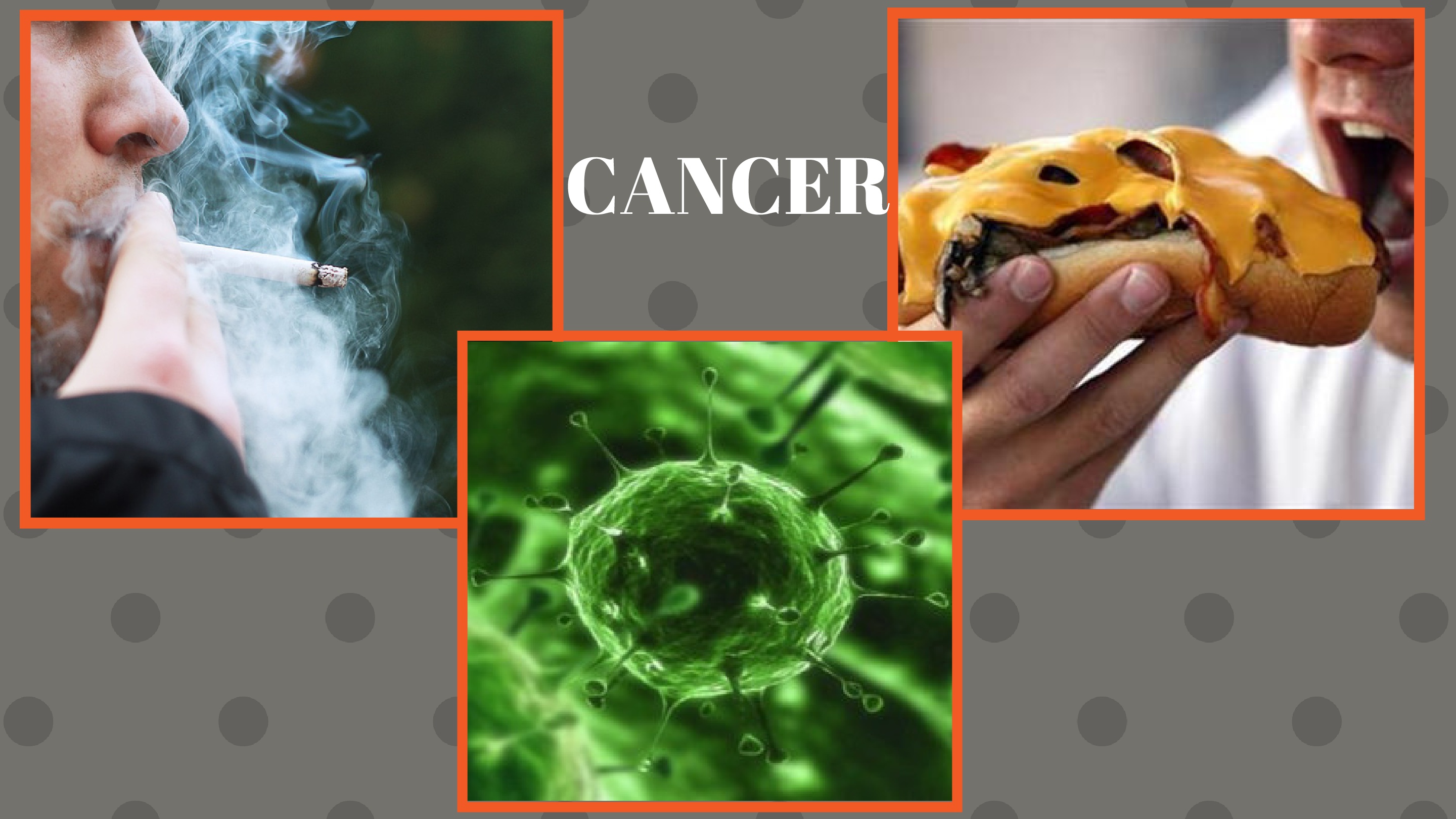
Being responsible for 13 percent or 8.2 million deaths worldwide, cancer bags the fifth position in the league of ‘Top 10 causes of death globally.’
Dismal figures color the sad picture of mortality due to cancer in India. With a total of 2.5 million Indians suffering from cancer, every 8 minutes a woman dies because of cervical cancer. Tobacco related diseases are alone responsible for 2,500 deaths daily. If 2 women are diagnosed of breast cancer, another woman would be dying because of it somewhere else.
For reference sake, let us see the ‘Top 10 causes of death globally’:
Ischemic heart disease
Stroke
Chronic obstructive pulmonary disease
Lower respiratory infections
Cancer
HIV & AIDS
Diarrhea
Diabetes
Road-traffic accidents
Hypertensive heart disease
Now if we observe carefully, these diseases have one thing in common – THESE CAUSES ARE LIFESTYLE RELATED. As much as there is to feel bad about these health problems, the good news is that most of these are PREVENTABLE; be it with a healthy diet, regular exercise, maintaining safety, or with periodic health check-ups.
Cancer is preventable too, provided steps are taken at the right time. Timely recognition of some ‘unnatural things’ happening with our bodies can help in preventing cancers at the primary level itself (at its time of onset).
Nobody intentionally asks for something bad to happen, but its always better to be on a safer side. We will go through various cancer detection tests and see which is suitable for which.
For convenience sake, we can study cancer detection under the following categories:
Pre-diagnostic concerns/Observations at home:
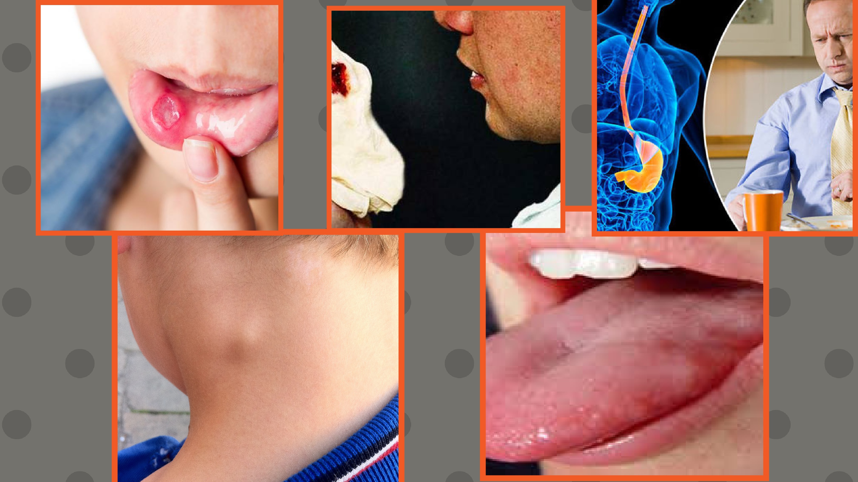
These are little ‘strange and different’ things we can observe in our bodies at home. Take a look –
Unexplained vaginal bleeding
A heartburn or indigestion problem that’s been bothering you for quite some time
Unexplained weight loss
An ulcer or sore that is taking too much time to heal or isn’t healing at all
Persistent coughs
A swelling or lump anywhere in the body
Blood showing up when coughing
A mole changing its size, color, etc.
Changes in size or shape of breasts
Difficult urination
Now before undergoing any diagnostic test, here are a few things one should keep in their mind:
You can prepare a set of questions to ask your doctor regarding any doubts about the test or treatment.
Do remember to inform the doctor about your medical history. It can be any disease, such as cardiovascular disorders, breathing problems, neurological disease, etc.
It is important that you let your doctor know about any present medications you are taking, be it antihypertensive drugs, pain meds, antidiabetic drugs, anything. If you are allergic to some material, tell them.
Most of the tests will require you to be on an empty stomach for at least a few hours before the procedure is carried out. Be sure to clear that out.
Diagnostic Tests:
Imaging
1. MRI Scan –
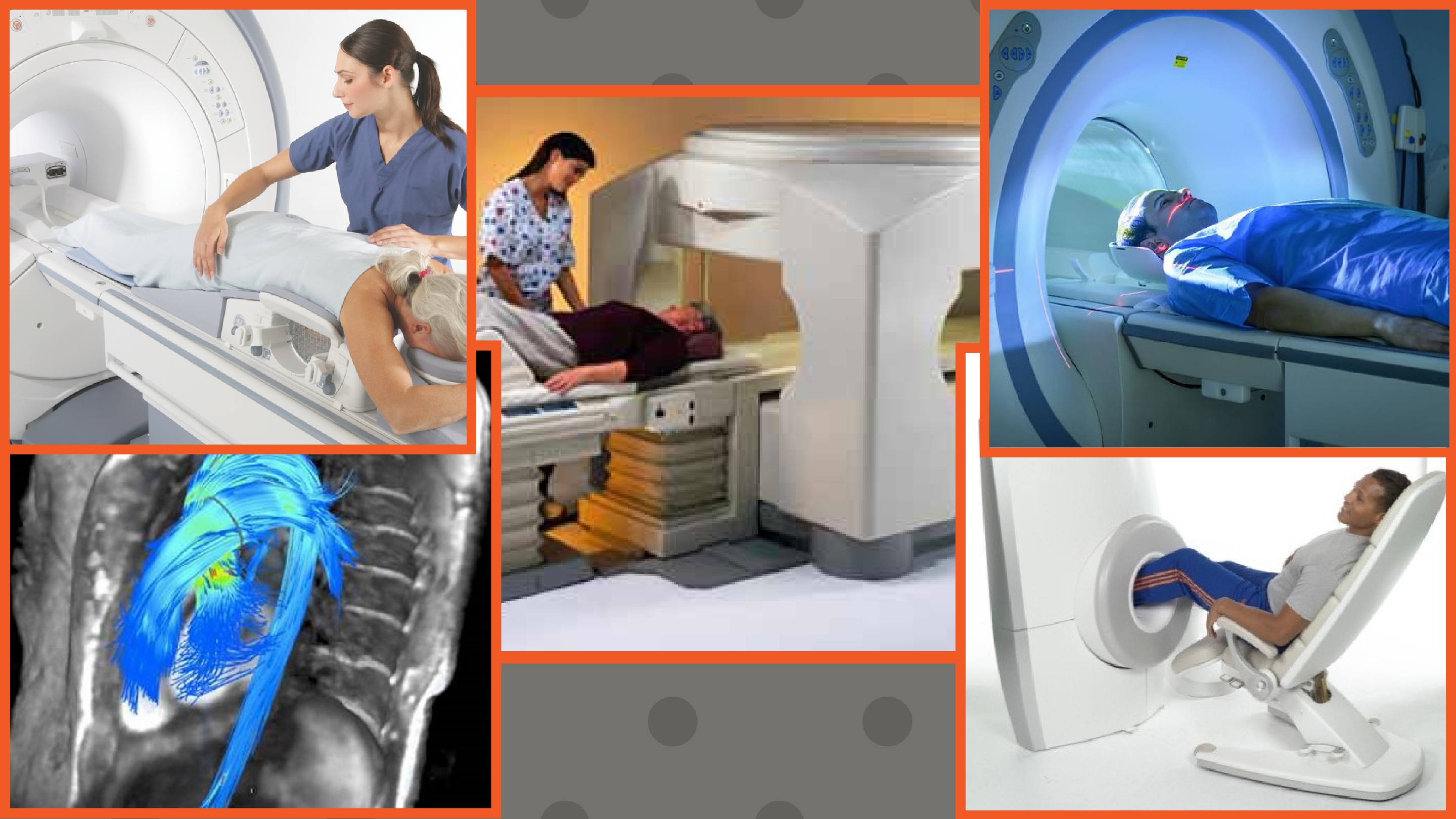
A magnetic resonance imaging scan uses magnetic waves and non ionizing radiation to create images of both hard and soft tissues of the body, including cancerous growths. With the help of an MRI, we can tell - The size and location of tumor.
Whether the tumor is localized or malignant.
If there are any chances of recurrence of tumor.
How to approach the treatment of tumor.
An MRI scan is carried out in a hollow cylindrical machine. You will be made to lie down on a sliding table which will slowly take you inside it for some time. It takes about 90 minutes to carry out the scan in which magnetic waves and radio waves create an image together. Due to the closed nature of an MRI machine, some people may feel claustrophobic inside it.
Different types of MRI scans are available; customized for different regions in the body such as:
- Breast MRI – Useful for detecting cancers, especially the ones with very dense, non-fatty breast tissue. It can locate tumors that a mammogram cannot. But, it cannot detect calcifications that a mammogram can. Hence, it must be treated only as an adjunct, not a substitute for a mammogram.
- Cardiac MRI (CMR) – It is a specialized technique where an MRI scan of the cardiac tissue is taken. An added benefit is that it allows the simultaneous recording of an ECG so that it can be compared with images taken in real time.
- Extremity MRI – This one is customized for use on the limbs (arms and legs mostly).
- Magnetic Resonance Angiography – MRI scan of the blood vessels can help in detection of abnormalities in circulation, along with vascular tumors.
- Neuroimaging – Helps in locating neoplastic growths in the brain. It can also be used in performing real time brain surgeries for removal of such growths.
2. Colonoscopy:
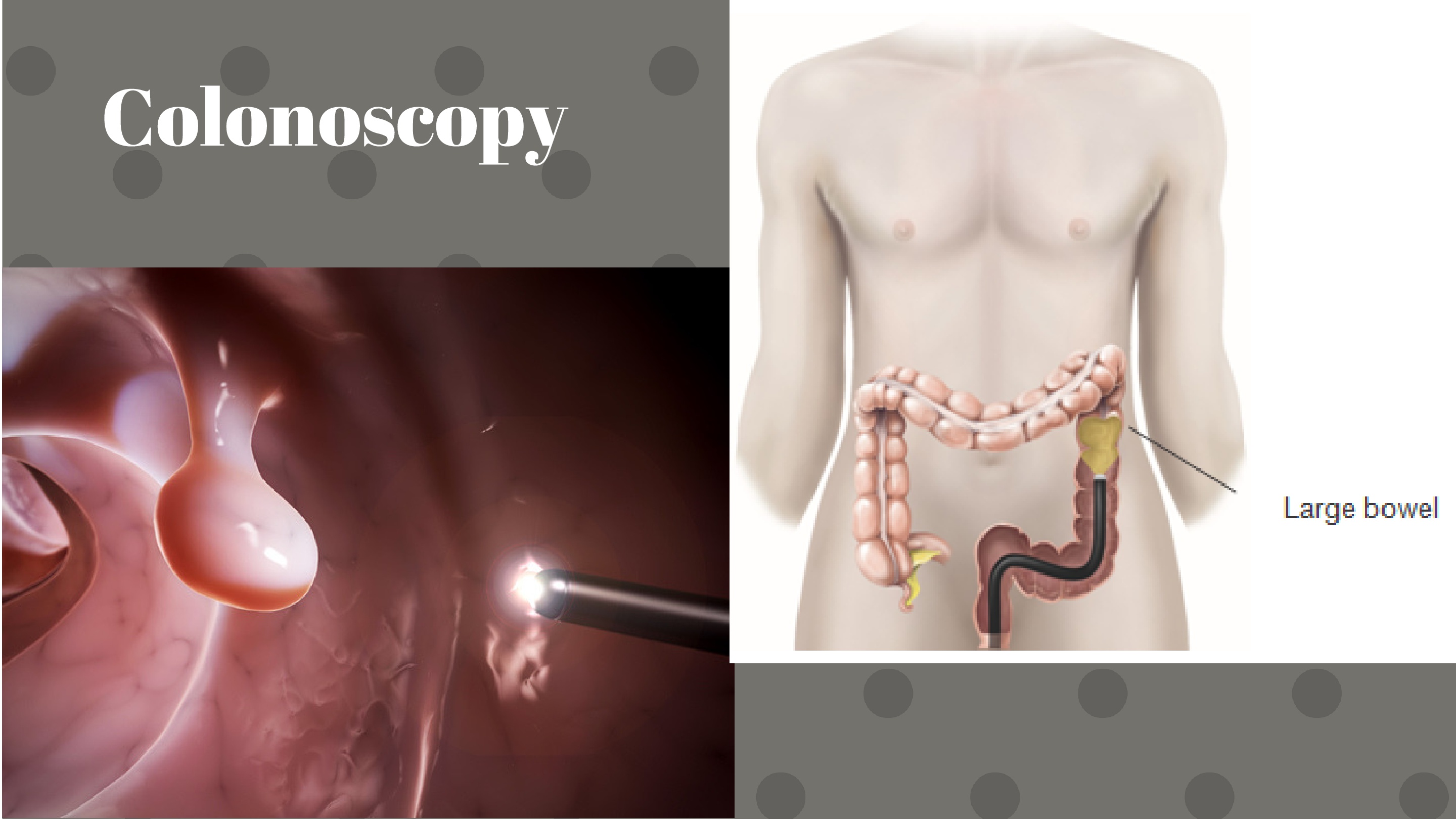
A colonoscope is a thin long flexible tube with the thickness of a finger and a tiny camera and a light source attached to its end. A gastroenterologist uses this device to view the condition of intestines and find out colorectal problems, if any. A sedative and a medication is given to reduce discomfort. The colonoscope is inserted via the anus into the colon. The camera shows the images on a monitor in the room where your doctor can see the condition of intestines. If needed, a biopsy of the suspected lesion is performed then and there for further histopathological examination.
3. Bone Scan: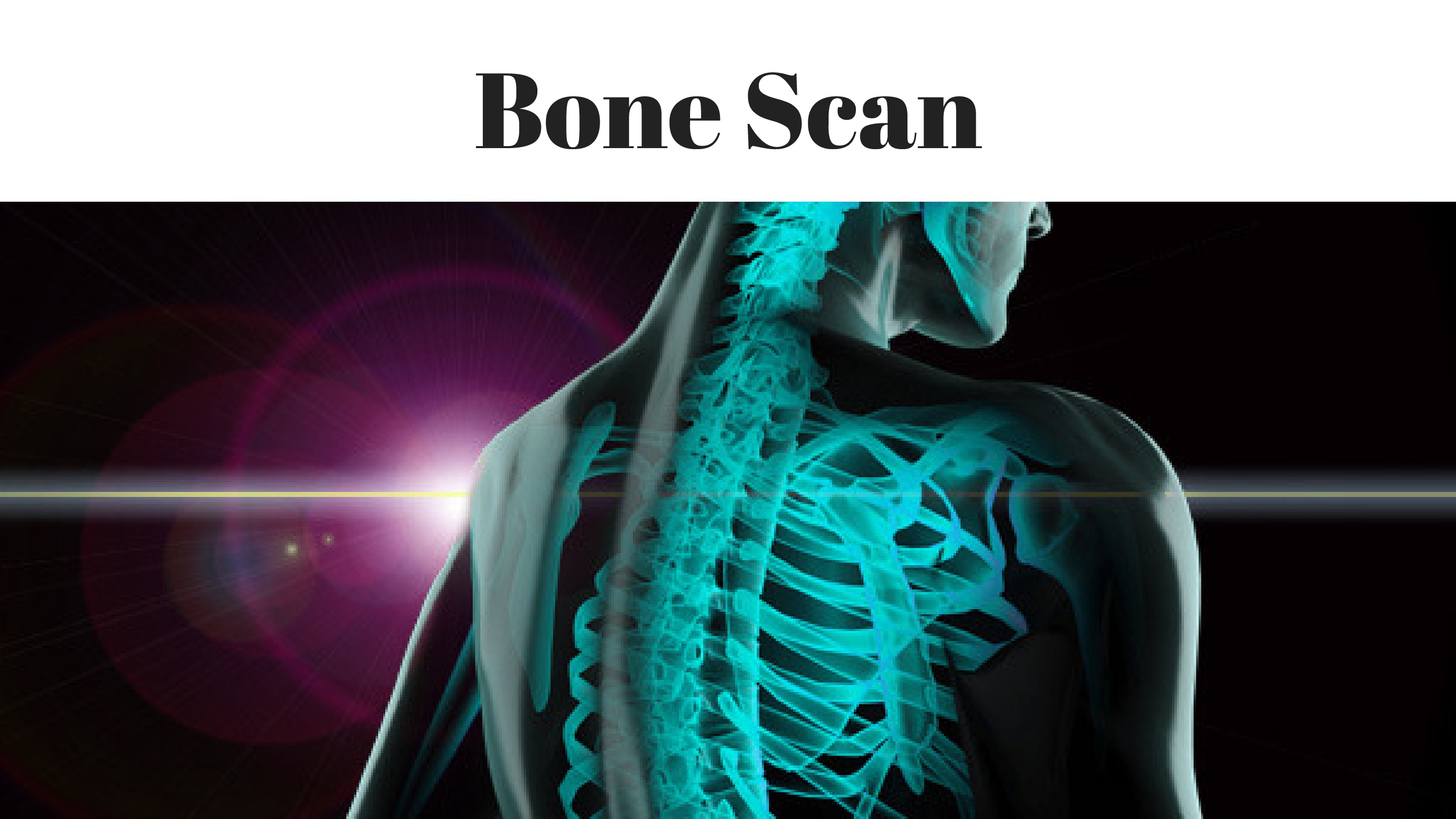
A bone scan is a type of nuclear medicine test. Here, small doses of radioactive material (technetium-99m), also known as a tracer, are inserted into the body via a vein. Once the tracer enters systemic circulation, it will spread inside and bind to the cells having cancer. If there are ‘bright/active’ areas seen on imaging, it indicates that deposits from primary tumors (e.g. lungs, breast, prostrate) are spreading to other parts in the body.
4. MUGA Scan:
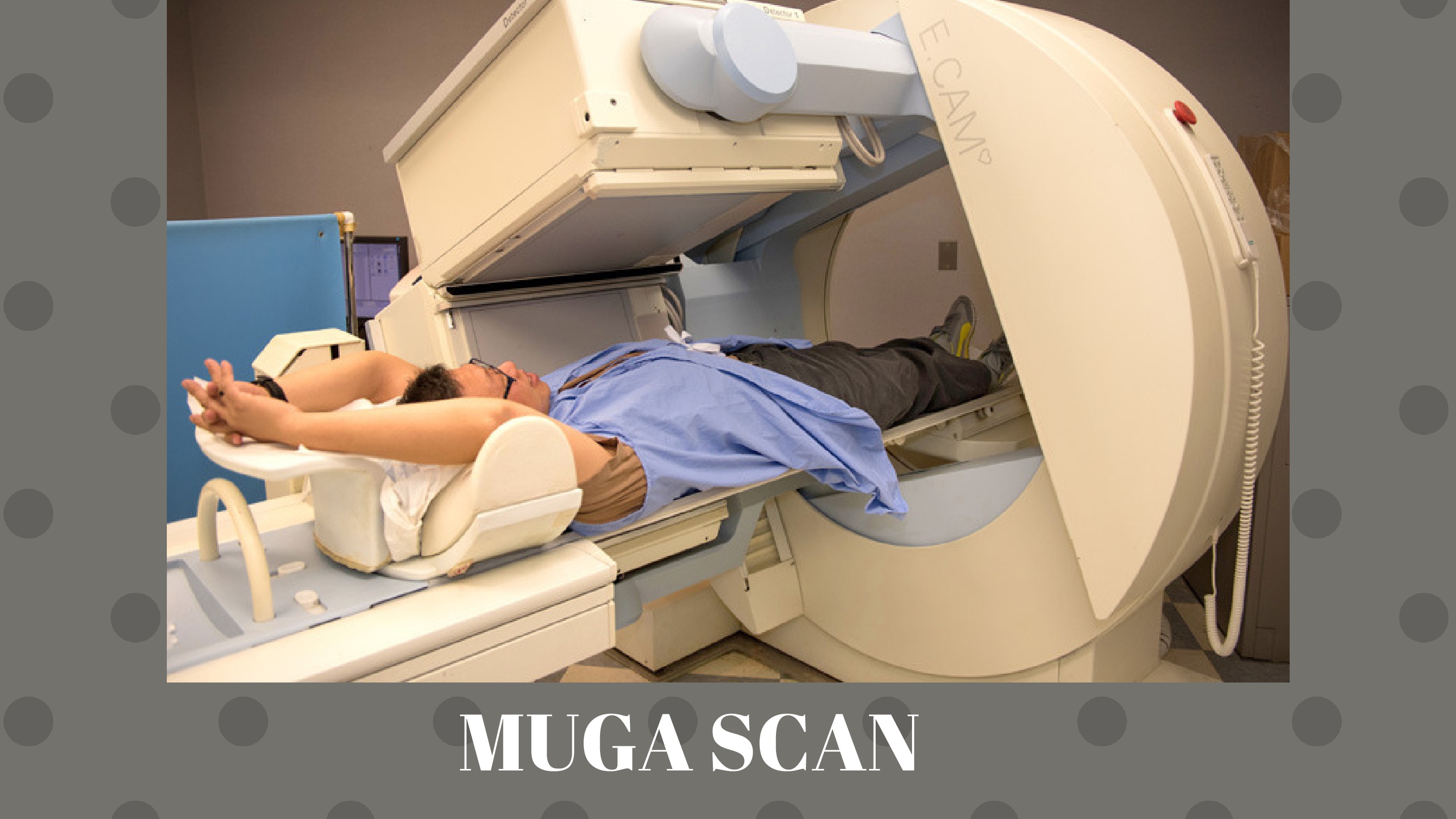
Also known as multigated acquisition scan, nuclear heart scan, radionuclide ventriculography or cardiac blood pooling imaging. A MUGA scan creates video images of the lower chambers of heart (ventricles) which helps the radiologist study their function. It can be used before providing chemotherapy to any cancer patient or when there is complaint of chest pain, dizziness, troubled breathing or tiredness.
Similar to a bone scan, radionuclide tracers are injected into the blood. A gamma camera now takes images of the heart at different time intervals.
Two types of scans are taken. One is while resting and the other is taken while exercising.
5. Computed Tomography (CT) Scan:
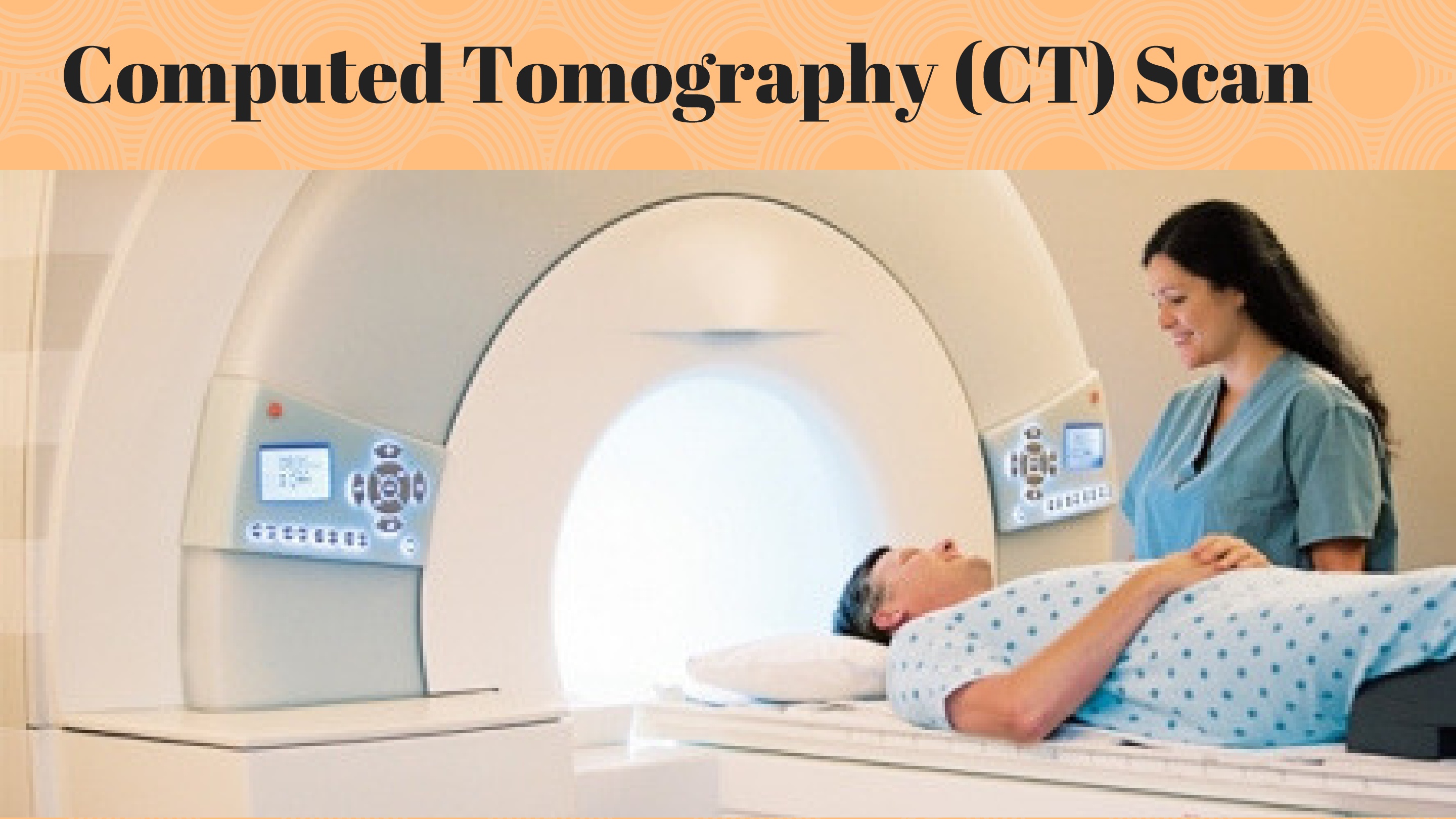
Also known to many as CAT scan, it uses X-rays to create a three dimensional image of the area of interest. Sometimes a contrast medium can be injected in the patient’s body in order to highlight a few areas.
The images will be recorded in sections and put together by a computer by a radiologist to create a complete picture. It is very useful in detecting tumors and other abnormalities.
6. PET-CT Scan:
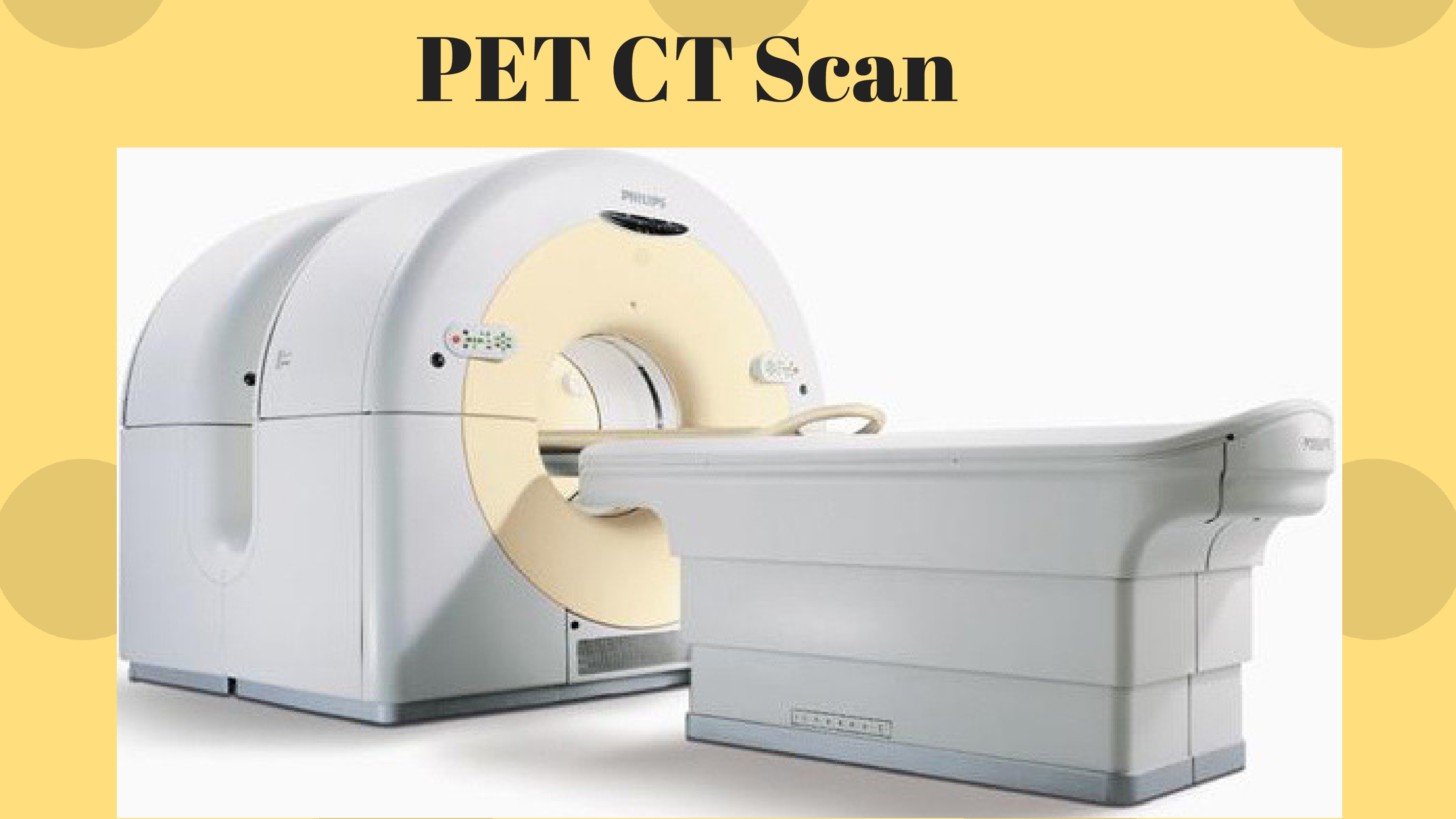
It is a very efficient diagnostic tool for catching cancers of the lung, lymph nodes, etc.
A PET-CT (Positron Emission Tomography) scan combines the metabolic tracing ability of a PET scan with 3D imaging of the CT scan. PET scan can detect metabolites (products of metabolism).
When it comes to detecting cancer, fluorine-18 or fluorodeoxyglucose is used as a tracer. Once injected in the vein, it is taken up by cells in the body (because the cells think it is glucose). Brain cells, cancer cells pick up glucose the most. Now these cells emit gamma rays which are picked up by the PET scan machine.
7. Mammography:
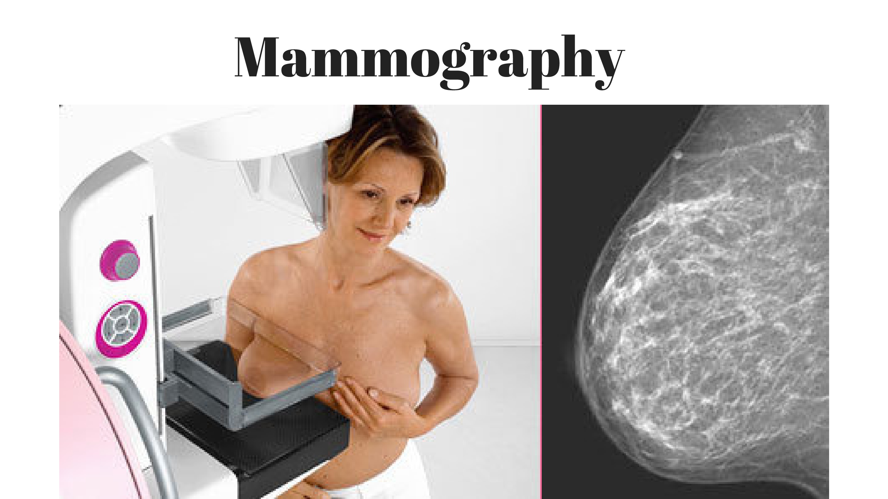
It is a picture of the breast created with the help of low dose X-rays. A mammogram is helpful; both in screening and diagnosing any breast abnormalities in women showing or not showing any particular symptoms.
It can also be used to examine the breast tissue in women who complain of pain, lump or any discharge from there. With advances in technology, mammography has evolved into –
- Digital mammography – X-rays of breast are converted into clearer digital images.
- Computer aided mammography – Uses its extra sensitivity to trace out areas of cancer that may have been ignored by a conventional mammogram.
As a general guideline, it is advisable that women who have entered their forties should go for a mammogram at least once a year.
8. Ultrasonography:
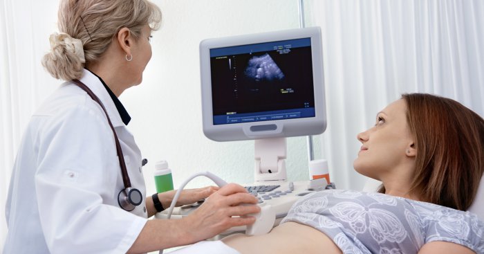
Ultrasonography uses the power of sound waves at high frequencies to help locating any suspected tumor or growth in the body. Ultrasonography also serves as a real time imaging guide while performing surgical procedures such as biopsies.
Sound waves can bounce off surfaces just like light. But their reflection depends on the surface they are reflecting from. This variation in bouncing of sound waves creates an echo, which is different for different organs. A transducer produces and captures these sound waves and converts them into an image which can be viewed on a monitor. Thus, abnormal tissues or masses can be differentiated from the normal ones.
9. Endoscopy:
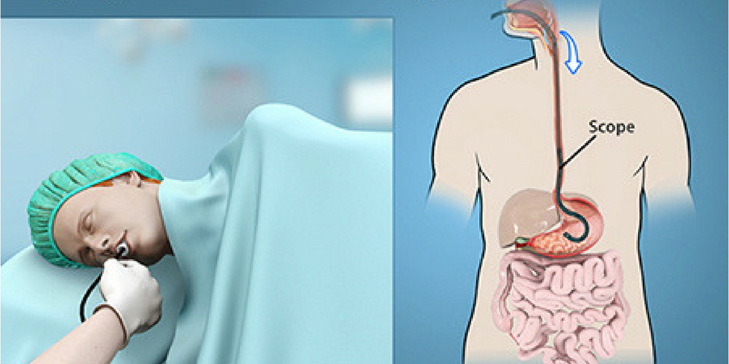
Endoscopy is examining a hollow cavity of the body by means of a camera tipped flexible tube called an endoscope. A tiny source of light on the endoscope helps in clear visualization once inside the body space (GIT, ear, throat, nasal cavity, etc.). Different types of endoscopy are available for different body parts, such as arthroscopy (for joints), colonoscopy (for intestines), sigmoidoscopy (for lower part of colon) etc.
Endoscopy benefits in the following ways –
Tissue samples can be collected using biopsy forceps.
Cytology smears can be prepared with brush samples collected by the endoscope.
Removal of sutures post-operatively.
10. Barium Enema:
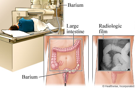
Enema means ‘injecting a fluid through rectum into the body.’ Barium is a radiopaque element i. e., it will show up as the brighter part on an X-ray. Hence, it acts as a contrast medium over here.
For 3 days, you will be asked to consume only liquids including fruit juices, fat-free soups, black coffee, etc. A laxative is given to be taken the day before procedure. These intend to clear your colon and rectum completely. On the day of procedure, you will be draped in a hospital gown. All metallic things are kept away. The radiologist will make you lie on the table and turn you on your side. After draping you with a cloth, a small tube is passed through your anus. Liquid barium will be administered in your body.
Once the radiologist is certain of the amount filled in, an X-ray will be taken. The bright white areas will show the areas barium has traveled. This helps in detecting suspected anomalies of the gastrointestinal tract such as diverticulitis, cancer, etc.
Laboratory diagnosis
1. Flow cytometry:
If one wants to define cell characteristics or properties (size, count, morphology, etc.), cytometry is the way to go. It can analyze multiple cell parameters in a fluid mixture at the same time. The cell population is ‘tagged’ or stained with a special fluorescent dye or antibodies and passed through a tube like device called a ‘cytometer.’ These cells are then passed under a laser beam. Now, as light strikes the cell surface, it will scatter the laser light. Based on this scattering property and other associated variables, cancer cells and their risk of recurrence can be measured. This is of particular interest in blood cancers such as leukemia or lymphomas.
2. Biopsy:
Biopsy is the examination of piece of living tissue to determine the cause, presence or extent of a disease. Biopsies are an important aid in cancer diagnosis. A tissue sample can be collected by performing a minor surgery or during any of the endoscopic tests if possible.
These tissue samples are then fixed, preserved and stained to be further studied under a microscope.
3. Cytology:
Cytology is the study of cells and their characteristics under a microscope. It is quick and easy way to detect abnormalities and requires minimum equipment and effort. That is why it is commonly used in epidemiological studies.
Cell samples are collected by any one of the following methods, stained and viewed under a microscope:
- Fine Needle Aspiration Cytology (FNAC) – A thin needle (23-25 gauge) is used to take out a small sample of the suspected tumor. Then the tissue is treated with stains and chemicals to understand the structure of cells present. This technique is of great value for large tumor masses, where an entire biopsy is not possible.
- Exfoliative Cytology – This is indicated in cases where the growth or lesion is not as suspicious. As the name suggests, cells which can be exfoliated (from lining epithelium) are collected with the help of wooden spatula or cytobrush, applied on a glass slide, fixed into place and then studied under a microscope.
- Abrasive Cytology – A very popular example of this technique is the ‘Pap smear,‘ used in the diagnosis of cervical cancer and evaluating precancerous lesions.
- Washings & Lavage – Normal saline is given and is reaspirated back. This way shed cells come out.
4. Histopathology:
It is the study of changes in tissue samples under a microscope. Tissues can be collected the help of surgery, biopsy or autopsy. The pathologist decides to preserve this section with the help of a technique known as ‘fixation.’ Tissue can be frozen or chemically fixed. After fixing, the prepared tissue slide is stained with chemicals such as hematoxylin-eosin stain, congo red, silver salts, oil red O, to allow easy viewing of different structures in the microscope.
5. Molecular diagnosis:
Cancer in its basic sense is a result of uncontrolled growth of cells. All cells contain a gene in them known as oncogene, which has the potential to cause cancer. They are expressed in high levels or mutated in neoplasms.
With the help of molecular diagnosis, mutations (alteration in gene structure) can be detected with the help of following methods:
- FISH – Fluorescent In Situ Hybridization is a test that ‘maps’ specific genes in the human cells. Colored dyes are attached to suspected parts on the chromosomes which are then studied under a fluorescent microscope. The target chromosome is studied for abnormalities in genetic structure (translocation, inversion, deletion or duplication).
FISH test is fast to produce results and can provide minute details that cannot be seen under a microscope.
- PCR – Polymerase Chain Reaction is a technique where small DNA fragments are copied and multiplied to a sufficient quantity on which studies can be done. DNA are generated in 30-40 PCR cycles.
PCR helps in detection of disease in a given DNA sample. Viral load can be detected too.
- Gene Chip Method – Here genes are scanned on oligonucleotide arrays called DNA chips, that can rapidly analyze thousands of genes at the same time.
6. Tumor markers:
Tumor markers are special biomarkers (glycoproteins, peptides, carbohydrates, enzymes, etc.) that are produced either by tumor cells or normal cells in response to presence of cancer. Some of these markers are produced in normal physiological conditions as well.
They can be used in staging, screening, monitoring progression, confirming diagnosis and determining the prognosis of cancer.
Tumor markers are detected in serum, less commonly in urine with help of immunoassay, enzyme activity studies or observing it in tissues using a microscope.
But it must be remembered that tumor markers are just an aid to cancer diagnosis, but not a confirmatory test in its own right.
7. Fecal Occult Blood Tests (FOBT):
As the name suggests, fecal occult blood refers to the blood hidden (or not readily visible) in the stools. This is of two types –
- Immunochemical FOBT – A tracer called an antibody attaches itself to the blood component of stools which can be easily detected.
- Guaiac FOBT – This test can be easily performed at home too. A card or pad is coated with guaiac. Once the stool sample is placed on this card, it will change color if any blood is present. This card is then sent either to the doctor or laboratory to calculate the results.
Apart from hemorrhoids (piles) and gastrointestinal bleeding, occult blood in stools may be indicative of colorectal cancer.
8. Immunocytochemistry & Immunohistochemistry:
These are tests which make use of special proteins in our body (antibodies) which are labeled. They bind the target cell after some time and then the distribution of this new product is studied to detect cancerous cells.
9. Digital Rectal Exam (DRE):
If someone constantly complains of bleeding via the rectum, abnormal changes in urine or bowel habits or urethral bleeding, then the doctor may suggest a DRE.
The lower rectum, belly and pelvis are checked in this test to screen for the following –
- Uterine, cervical or ovarian cancer
- Rectal cancer
- Lower colon cancer
- Prostate cancer
A DRE is done as part of a routine medical exam. It is carried out in a lying down position with the legs either raised or held close to the chest.
Thanks to economical technology, most of these cancer screening tests are available in majority of diagnostic centers. Some of these tests have potential to reduce the risk of cancer, while some have been deemed as a ‘secondary choice’ to cancer diagnosis. It is therefore useful to discuss with your doctor the importance of each test before going ahead.
Tests like mammography, pap smear, human papillomavirus (HPV) detection, colonoscopy etc. have proved themselves useful in reducing the risk of death due to cancer.
India is among the few countries which has its own National Cancer Control Programme. It emphasizes on early diagnosis, treatment of cancer and distribution of appropriate therapy among the general population.
Tobacco related cancers, uterine & cervical cancers top the priority list.
Thus, it is possible that with modification of lifestyle (controlling alcohol, tobacco, obesity, inactivity) and regular check-ups, cancer can be caught at an early stage and dealt with efficiently. Now the next time you visit your doctor, take the initiative to discuss about the various risks of cancer and measures to prevent them.
The journey to better health begins with a single step after all.